ID:CAC1S_HUMAN DESCRIPTION: RecName: Full=Voltage-dependent L-type calcium channel subunit alpha-1S; AltName: Full=Calcium channel, L type, alpha-1 polypeptide, isoform 3, skeletal muscle; AltName: Full=Voltage-gated calcium channel subunit alpha Cav1.1; FUNCTION: Voltage-sensitive calcium channels (VSCC) mediate the entry of calcium ions into excitable cells and are also involved in a variety of calcium-dependent processes, including muscle contraction, hormone or neurotransmitter release, gene expression, cell motility, cell division and cell death. The isoform alpha-1S gives rise to L-type calcium currents. Long-lasting (L-type) calcium channels belong to the 'high-voltage activated' (HVA) group. They are blocked by dihydropyridines (DHP), phenylalkylamines, benzothiazepines, and by omega-agatoxin-IIIA (omega-Aga-IIIA). They are however insensitive to omega-conotoxin- GVIA (omega-CTx-GVIA) and omega-agatoxin-IVA (omega-Aga-IVA). Calcium channels containing the alpha-1S subunit play an important role in excitation-contraction coupling in skeletal muscle. SUBUNIT: Multisubunit complex consisting of alpha-1, alpha-2, beta and delta subunits in a 1:1:1:1 ratio. The channel activity is directed by the pore-forming and voltage-sensitive alpha-1 subunit. In many cases, this subunit is sufficient to generate voltage-sensitive calcium channel activity. The auxiliary subunits beta and alpha-2/delta linked by a disulfide bridge regulate the channel activity. An additional gamma subunit is present only in skeletal muscle L-type channel. Interacts with DYSF and JSRP1. Interacts with RYR1 (By similarity). SUBCELLULAR LOCATION: Membrane; Multi-pass membrane protein. TISSUE SPECIFICITY: Skeletal muscle specific. DOMAIN: Each of the four internal repeats contains five hydrophobic transmembrane segments (S1, S2, S3, S5, S6) and one positively charged transmembrane segment (S4). S4 segments probably represent the voltage-sensor and are characterized by a series of positively charged amino acids at every third position. DOMAIN: The loop between repeats II and III interacts with the ryanodine receptor, and is therefore important for calcium release from the endoplasmic reticulum necessary for muscle contraction. PTM: Phosphorylation by PKA activates the calcium channel (By similarity). DISEASE: Defects in CACNA1S are the cause of periodic paralysis hypokalemic type 1 (HOKPP1) [MIM:170400]; also designated HYPOPP. HOKPP1 is an autosomal dominant disorder manifested by episodic flaccid generalized muscle weakness associated with falls of serum potassium levels. DISEASE: Genetic variations in CACNA1S are the cause of susceptibility to malignant hyperthermia 5 (MHS5) [MIM:601887]; an autosomal dominant disorder that is potentially lethal in susceptible individuals on exposure to commonly used inhalational anesthetics and depolarizing muscle relaxants. DISEASE: Defects in CACNA1S are the cause of susceptibility to thyrotoxic periodic paralysis type 1 (TTPP1) [MIM:188580]. A sporadic muscular disorder characterized by episodic weakness and hypokalemia during a thyrotoxic state. It is clinically similar to hereditary hypokalemic periodic paralysis, except for the fact that hyperthyroidism is an absolute requirement for disease manifestation. The disease presents with recurrent episodes of acute muscular weakness of the four extremities that vary in severity from paresis to complete paralysis. Attacks are triggered by ingestion of a high carbohydrate load or strenuous physical activity followed by a period of rest. Thyrotoxic periodic paralysis can occur in association with any cause of hyperthyroidism, but is most commonly associated with Graves disease. SIMILARITY: Belongs to the calcium channel alpha-1 subunit (TC 1.A.1.11) family. CACNA1S subfamily. WEB RESOURCE: Name=GeneReviews; URL="http://www.ncbi.nlm.nih.gov/sites/GeneTests/lab/gene/CACNA1S";
The RNAfold program from the Vienna RNA Package is used to perform the secondary structure predictions and folding calculations. The estimated folding energy is in kcal/mol. The more negative the energy, the more secondary structure the RNA is likely to have.
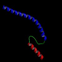
ModBase Predicted Comparative 3D Structure on Q13698
Front
Top
Side
The pictures above may be empty if there is no ModBase structure for the protein. The ModBase structure frequently covers just a fragment of the protein. You may be asked to log onto ModBase the first time you click on the pictures. It is simplest after logging in to just click on the picture again to get to the specific info on that model.
Orthologous Genes in Other Species
Orthologies between human, mouse, and rat are computed by taking the best BLASTP hit, and filtering out non-syntenic hits. For more distant species reciprocal-best BLASTP hits are used. Note that the absence of an ortholog in the table below may reflect incomplete annotations in the other species rather than a true absence of the orthologous gene.
Gene Ontology (GO) Annotations with Structured Vocabulary
Molecular Function: GO:0005216 ion channel activity GO:0005244 voltage-gated ion channel activity GO:0005245 voltage-gated calcium channel activity GO:0005262 calcium channel activity GO:0005515 protein binding GO:0005516 calmodulin binding GO:0008331 high voltage-gated calcium channel activity GO:0046872 metal ion binding
Biological Process: GO:0006811 ion transport GO:0006816 calcium ion transport GO:0006936 muscle contraction GO:0034765 regulation of ion transmembrane transport GO:0055085 transmembrane transport GO:0070588 calcium ion transmembrane transport GO:0071313 cellular response to caffeine GO:0086010 membrane depolarization during action potential
 Sequence and Links to Tools and Databases
  Common Gene Haplotype Alleles