ID:CA2D4_HUMAN DESCRIPTION: RecName: Full=Voltage-dependent calcium channel subunit alpha-2/delta-4; AltName: Full=Voltage-gated calcium channel subunit alpha-2/delta-4; Contains: RecName: Full=Voltage-dependent calcium channel subunit alpha-2-4; Contains: RecName: Full=Voltage-dependent calcium channel subunit delta-4; Flags: Precursor; FUNCTION: The alpha-2/delta subunit of voltage-dependent calcium channels regulates calcium current density and activation/inactivation kinetics of the calcium channel. SUBUNIT: Dimer formed of alpha-2-2 and delta-2 chains; disulfide- linked. Voltage-dependent calcium channels are multisubunit complexes, consisting of alpha-1 (CACNA1), alpha-2 (CACNA2D), beta (CACNB) and delta (CACNA2D) subunits in a 1:1:1:1 ratio (Probable). Interacts with CACNA1C and CACNB3. SUBCELLULAR LOCATION: Membrane; Single-pass type I membrane protein (Potential). TISSUE SPECIFICITY: Predominantly expressed in certain types of endocrine cells. Present in the Paneth cells of the small intestine. Also present in the erythroblasts in the fetal liver, in the cells of the zona reticularis of the adrenal gland and in the basophils of the pituitary. Present at low level in some brain regions such as the cerebellum (at protein level). DOMAIN: The MIDAS-like motif in the VWFA domain binds divalent metal cations and is required to promote trafficking of the alpha- 1 (CACNA1) subunit to the plasma membrane by an integrin-like switch (By similarity). PTM: May be proteolytically processed into subunits alpha-2-4 and delta-4 that are disulfide-linked. It is however unclear whether such cleavage really takes place in vivo and has a functional role (By similarity). DISEASE: Defects in CACNA2D4 are the cause of retinal cone dystrophy 4 (RCD4) [MIM:610478]. RCD4 is characterized by minimal symptoms except for slowly progressive reduction in visual acuity. MISCELLANEOUS: In contrast to CACNA2D1 and CACNA2D2, it does not bind gabapentin, an antiepileptic drug. SIMILARITY: Belongs to the calcium channel subunit alpha-2/delta family. SIMILARITY: Contains 1 cache domain. SIMILARITY: Contains 1 VWFA domain. SEQUENCE CAUTION: Sequence=AAH48288.1; Type=Erroneous translation; Note=Wrong choice of frame; Sequence=AAN06672.1; Type=Erroneous initiation; Note=Translation N-terminally extended;
The RNAfold program from the Vienna RNA Package is used to perform the secondary structure predictions and folding calculations. The estimated folding energy is in kcal/mol. The more negative the energy, the more secondary structure the RNA is likely to have.
Pfam Domains: PF00092 - von Willebrand factor type A domain PF08399 - VWA N-terminal PF08473 - Neuronal voltage-dependent calcium channel alpha 2acd PF13519 - von Willebrand factor type A domain PF13768 - von Willebrand factor type A domain
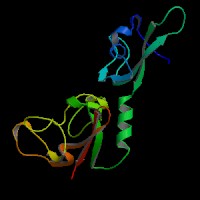
ModBase Predicted Comparative 3D Structure on Q7Z3S7
Front
Top
Side
The pictures above may be empty if there is no ModBase structure for the protein. The ModBase structure frequently covers just a fragment of the protein. You may be asked to log onto ModBase the first time you click on the pictures. It is simplest after logging in to just click on the picture again to get to the specific info on that model.
Orthologous Genes in Other Species
Orthologies between human, mouse, and rat are computed by taking the best BLASTP hit, and filtering out non-syntenic hits. For more distant species reciprocal-best BLASTP hits are used. Note that the absence of an ortholog in the table below may reflect incomplete annotations in the other species rather than a true absence of the orthologous gene.
Gene Ontology (GO) Annotations with Structured Vocabulary
Molecular Function: GO:0005244 voltage-gated ion channel activity GO:0005245 voltage-gated calcium channel activity GO:0005262 calcium channel activity GO:0046872 metal ion binding
Biological Process: GO:0006811 ion transport GO:0006816 calcium ion transport GO:0034765 regulation of ion transmembrane transport GO:0050908 detection of light stimulus involved in visual perception GO:0070588 calcium ion transmembrane transport
 Sequence and Links to Tools and Databases
  Common Gene Haplotype Alleles