ID:CAD23_HUMAN DESCRIPTION: RecName: Full=Cadherin-23; AltName: Full=Otocadherin; Flags: Precursor; FUNCTION: Cadherins are calcium-dependent cell adhesion proteins. They preferentially interact with themselves in a homophilic manner in connecting cells. CDH23 is required for establishing and/or maintaining the proper organization of the stereocilia bundle of hair cells in the cochlea and the vestibule during late embryonic/early postnatal development. It is part of the functional network formed by USH1C, USH1G, CDH23 and MYO7A that mediates mechanotransduction in cochlear hair cells. Required for normal hearing. SUBUNIT: Interacts with PCDH15 (By similarity). Interacts with USH1C and USH1G. SUBCELLULAR LOCATION: Cell membrane; Single-pass type I membrane protein (By similarity). TISSUE SPECIFICITY: Particularly strong expression in the retina. Found also in the cochlea. DISEASE: Defects in CDH23 are the cause of Usher syndrome type 1D (USH1D) [MIM:601067]. USH is a genetically heterogeneous condition characterized by the association of retinitis pigmentosa and sensorineural deafness. Age at onset and differences in auditory and vestibular function distinguish Usher syndrome type 1 (USH1), Usher syndrome type 2 (USH2) and Usher syndrome type 3 (USH3). USH1 is characterized by profound congenital sensorineural deafness, absent vestibular function and prepubertal onset of progressive retinitis pigmentosa leading to blindness. DISEASE: Defects in CDH23 are a cause of Usher syndrome type 1D/F (USH1DF) [MIM:601067]. USH1DF patients are heterozygous for mutations in CDH23 and PCDH15, indicating a digenic inheritance pattern. DISEASE: Defects in CDH23 are the cause of deafness autosomal recessive type 12 (DFNB12) [MIM:601386]. DFNB12 is a form of sensorineural hearing loss. Sensorineural deafness results from damage to the neural receptors of the inner ear, the nerve pathways to the brain, or the area of the brain that receives sound information. SIMILARITY: Contains 27 cadherin domains. WEB RESOURCE: Name=Mutations of the CDH23 gene; Note=Retina International's Scientific Newsletter; URL="http://www.retina-international.org/files/sci-news/cdh23mut.htm"; WEB RESOURCE: Name=GeneReviews; URL="http://www.ncbi.nlm.nih.gov/sites/GeneTests/lab/gene/CDH23";
The RNAfold program from the Vienna RNA Package is used to perform the secondary structure predictions and folding calculations. The estimated folding energy is in kcal/mol. The more negative the energy, the more secondary structure the RNA is likely to have.
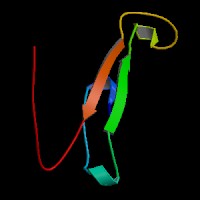
ModBase Predicted Comparative 3D Structure on Q9H251
Front
Top
Side
The pictures above may be empty if there is no ModBase structure for the protein. The ModBase structure frequently covers just a fragment of the protein. You may be asked to log onto ModBase the first time you click on the pictures. It is simplest after logging in to just click on the picture again to get to the specific info on that model.
Orthologous Genes in Other Species
Orthologies between human, mouse, and rat are computed by taking the best BLASTP hit, and filtering out non-syntenic hits. For more distant species reciprocal-best BLASTP hits are used. Note that the absence of an ortholog in the table below may reflect incomplete annotations in the other species rather than a true absence of the orthologous gene.
 Sequence and Links to Tools and Databases
  Common Gene Haplotype Alleles