ID:HTRA1_HUMAN DESCRIPTION: RecName: Full=Serine protease HTRA1; EC=3.4.21.-; AltName: Full=High-temperature requirement A serine peptidase 1; AltName: Full=L56; AltName: Full=Serine protease 11; Flags: Precursor; FUNCTION: Serine protease with a variety of targets, including extracellular matrix proteins such as fibronectin. HTRA1-generated fibronectin fragments further induce synovial cells to up-regulate MMP1 and MMP3 production. May also degrade proteoglycans, such as aggrecan, decorin and fibromodulin. Through cleavage of proteoglycans, may release soluble FGF-glycosaminoglycan complexes that promote the range and intensity of FGF signals in the extracellular space. Regulates the availability of insulin-like growth factors (IGFs) by cleaving IGF-binding proteins. Inhibits signaling mediated by TGF-beta family members. This activity requires the integrity of the catalytic site, although it is unclear whether TGF-beta proteins are themselves degraded. By acting on TGF-beta signaling, may regulate many physiological processes, including retinal angiogenesis and neuronal survival and maturation during development. Intracellularly, degrades TSC2, leading to the activation of TSC2 downstream targets. SUBUNIT: Forms homotrimers. In the presence of substrate, may form higher-order multimers in a PDZ-independent manner. Interacts with TGF-beta family members, including BMP4, TGFB1, TGFB2, activin A and GDF5 (By similarity). SUBCELLULAR LOCATION: Secreted. Cytoplasm, cytosol. Note=Predominantly secreted. Also found associated with the plasma membrane. TISSUE SPECIFICITY: Widely expressed, with strongest expression in placenta (at protein level). Secreted by synovial fibroblasts. Up- regulated in osteoarthritis and rheumatoid arthritis synovial fluids and cartilage as compared with non-arthritic (at protein level). DEVELOPMENTAL STAGE: In the placenta, in the first trimester of gestation, low expression in the cells surrounding villi both in the inner layer of the cytotrophoblast and in the outer layer of the syncytiotrophoblast (at protein level). In the third trimester of gestation, very strong expression in the outer layer forming the syncytiotrophoblast and lower in the cytotrophoblast (at protein level). DOMAIN: The IGFBP N-terminal domain mediates interaction with TSC2 substrate. DISEASE: Variations in the promoter region of HTRA1 are the cause of susceptibility to age-related macular degeneration type 7 (ARMD7) [MIM:610149]. ARMD is the leading cause of vision loss and blindness among older individuals in the developed word. It is classified as either dry (nonneovascular) or wet (neovascular). ARMD7 is a wet form, in which new blood vessels form and break beneath the retina. This leakage causes permanent damage to surrounding retinal tissue, distorting and destroying central vision. Wet ARMD is more prevalent among Asians than Caucasians. DISEASE: Defects in HTRA1 are the cause of cerebral autosomal recessive arteriopathy with subcortical infarcts and leukoencephalopathy (CARASIL) [MIM:600142]. CARASIL is characterized by nonhypertensive cerebral small-vessel arteriopathy with subcortical infarcts, alopecia, and spondylosis, with an onset in early adulthood. On neuropathological examination, atherosclerosis associated with intimal thickening and dense collagen fibers, loss of vascular smooth-muscle cells, and hyaline degeneration of the tunica media has been observed in cerebral small arteries. SIMILARITY: Belongs to the peptidase S1B family. SIMILARITY: Contains 1 IGFBP N-terminal domain. SIMILARITY: Contains 1 Kazal-like domain. SIMILARITY: Contains 1 PDZ (DHR) domain. WEB RESOURCE: Name=GeneReviews; URL="http://www.ncbi.nlm.nih.gov/sites/GeneTests/lab/gene/HTRA1";
The RNAfold program from the Vienna RNA Package is used to perform the secondary structure predictions and folding calculations. The estimated folding energy is in kcal/mol. The more negative the energy, the more secondary structure the RNA is likely to have.
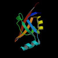
ModBase Predicted Comparative 3D Structure on Q92743
Front
Top
Side
The pictures above may be empty if there is no ModBase structure for the protein. The ModBase structure frequently covers just a fragment of the protein. You may be asked to log onto ModBase the first time you click on the pictures. It is simplest after logging in to just click on the picture again to get to the specific info on that model.
Orthologous Genes in Other Species
Orthologies between human, mouse, and rat are computed by taking the best BLASTP hit, and filtering out non-syntenic hits. For more distant species reciprocal-best BLASTP hits are used. Note that the absence of an ortholog in the table below may reflect incomplete annotations in the other species rather than a true absence of the orthologous gene.
 Sequence and Links to Tools and Databases
  Common Gene Haplotype Alleles